
Your femur gives you a leg to stand on, literally.

Use your cane or walker if you have difficulty walking or have an increased risk for falls.Follow a diet and exercise plan that will help you maintain good bone health.Never stand on chairs, tables or countertops. Always use the proper tools or equipment at home to reach things.
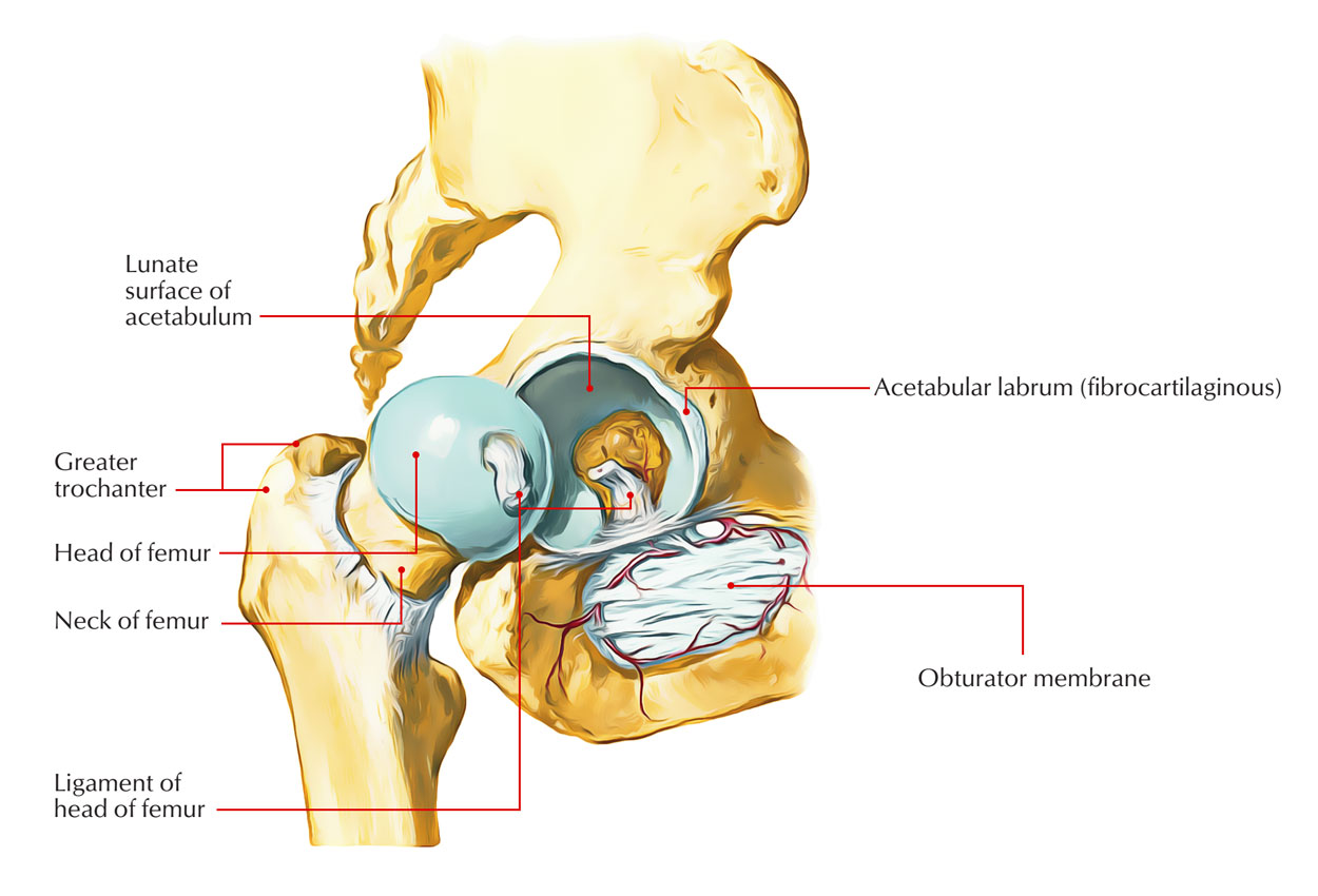
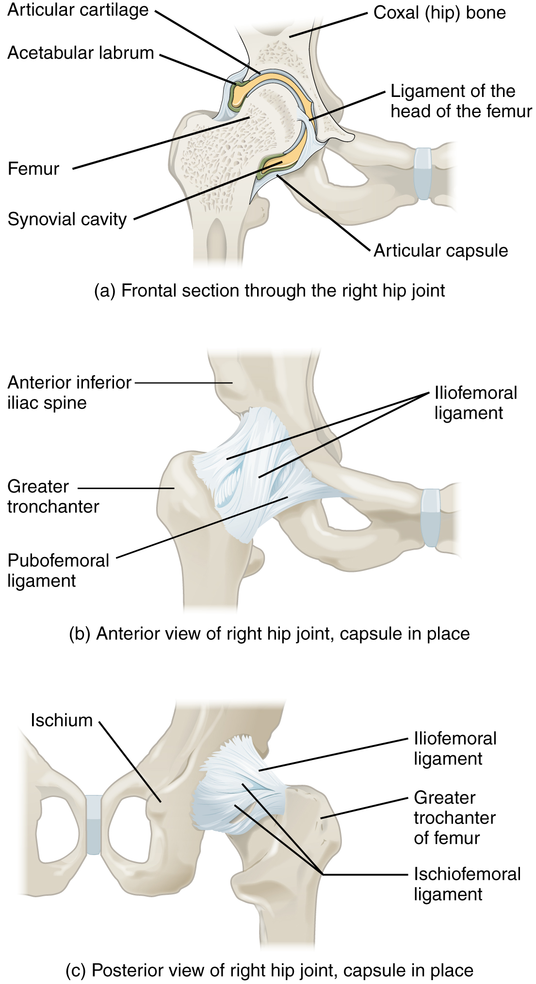
COMPLETE ANATOMY OF HIP JOINT FREE

It’s sometimes called runner’s or jumper’s knee. Patellofemoral pain syndrome (PFPS) is pain around and under your kneecap (patella). Talk to your provider about a bone density screening that can catch osteoporosis before it causes a fracture. Women, people assigned female at birth and adults older than 50 have an increased risk for developing osteoporosis. Many people don’t know they have osteoporosis until after it causes them to break a bone. Osteoporosis weakens bones, making them more susceptible to sudden and unexpected fractures. Go to the emergency room right away if you’ve experienced a trauma or think you have a fracture.

A deformity or bump that’s not usually on your body.Inability to move your leg like you usually can.Because femurs are so strong, they’re usually only broken by serious injuries like car accidents, falls or other traumas. Femur fracturesĪ bone fracture is the medical term for breaking a bone. The most common issues that affect femurs are fractures, osteoporosis and patellofemoral pain syndrome. What are the common conditions and disorders that affect the femur? It can support as much as 30 times the weight of your body. The femur is also the strongest bone in your body. Most adult femurs are around 18 inches long. Your femur is the largest bone in your body. If you ever break your femur - a femoral fracture - your provider might use some of these terms to describe where your bone was damaged. It includes the:Īll of these parts and labels are usually more for your healthcare provider to use as they describe where you’re having pain or issues. It meets your tibia (shin) and patella (kneecap). The lower (distal) end of your femur forms the top of your knee joint. It angles slightly toward the center of your body. The shaft is the long portion of the femur that supports your weight and forms the structure of your thigh. The upper (proximal) end of your femur connects to your hip joint. It’s the classic shape used for bones in cartoons: A cylinder with two round bumps at each end.Įven though it’s one long bone, your femur is made up of several parts. The femur has two rounded ends and a long shaft in the middle. The femur is the only bone in your thigh.


 0 kommentar(er)
